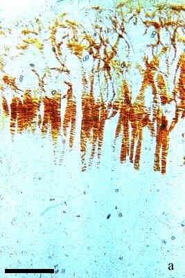
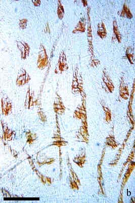
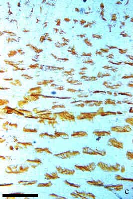
Fig.4. Patterns of the corneal coloration in Irish lord,
Hemilepidotus gilberti at dark adapted (a),
semi-adapted (b) and light adapted (c) conditions.
All slides show the dorsal part of the cornea. Scale bar = 500 mkm.
Almost all pigment is located in cell bodies and adjacent
parts of processes;
Pigment moves to distal expanded ends of the processes;
All pigment is located in the end expansions of cell processes,
no pigmented parts of processes are visible.
Back to the Poster Main Page