| species | activity | retina | dopaminergic neurons |
|---|---|---|---|
| crucian carp |   |

 | |
| scad pickarel red mullet |  |
dominated | 
 |
| whiting | |
dominated | 
 |
| frogs | |
| |
| gecko | |
 | |
| lizard | |
| |
| grass-snake |
|
|
 A paper version of the poster was presented at the 24th Goettingen Neurobiology Conference
on Brain and Evolution, May 31 - June 2, 1996.
A paper version of the poster was presented at the 24th Goettingen Neurobiology Conference
on Brain and Evolution, May 31 - June 2, 1996.
Poster No 372.
Presentation time - from Friday noon until Saturday noon.
Published in:
N.Elsner ed., Proceedings of the 24th Goettingen Neurobiology Conference
on Brain and Evolution, Volume II, Thieme Verlag, Stuttgart, 1996
 Dopaminergic amacrine cells in mammals are known to play an essential
role in switching the scotopic vision to photopic one. The number
of these cells correlates with number of rods.
Dopaminergic amacrine cells in mammals are known to play an essential
role in switching the scotopic vision to photopic one. The number
of these cells correlates with number of rods.
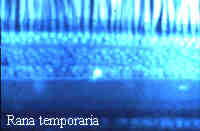
We studied dopaminergic cells in cold-blooded animals with different types of activity and photoreceptor contents of the retinas: marine (scad Trachurus mediterraneus ponticus, pickarel Spicara smaris, red mullet Mullus barbatus ponticus and whiting Merlangius merlangus euxinus) and freshwater (crucian carp Carassius carassius) fishes, frogs Rana temporaria and R. ridibunda, gecko Gymnodactylus caspius, diurnal lizard Phrynocephalus mystaceus and grass-snake Natrix natrix. Dopaminergic neurons were visualized by FaGlu method or stained with antiserum against TH. Their morphology was investigated in flatmounted retinae and radial sections.
| species | activity | retina | dopaminergic neurons |
|---|---|---|---|
| crucian carp | |
| |
| scad pickarel red mullet | |
dominated |
|
| whiting | |
dominated |
|
| frogs | |
| |
| gecko | |
| |
| lizard | |
| |
| grass-snake |
|
|
In the whiting, predominantly rod, retina multiple monostratified amacrines were found. Bistratified amacrines were found in predominantly cone retinas of some perciformes. Only interplexiform (bistratified in IPL) dopaminergic cells exist in crucian carp mixed retina. Frogs that are active all the day round and have mixed retinae were found to posses both mono- and bistratified amacrines. In the cone retina of the grass-snake there are the same two types of dopaminergic amacrine cells. Similar picture was observed in the all-cone retina of the caspian gecko. Interplexiform cells were found there too. In the cone retina of diurnal lizard Phrynocephalus mystaceus numerous exclusively bistratified dopaminergic amacrines were found. The presence of mono- or bistratified dopaminergic amacrines in the retina appears to correlate rather with the type of activity than with the predominance of cones or rods in the retina.
E.M.Maximova, I.A.Utina. Dopamine-containing cells in the caspian gecko retina. Sensory Systems, 4(1):21-24, 1990 (in Russian).
E.M.Maximova, I.A.Utina. Bistratified dopaminergic amacrine cells in the retinae of lower vertebrates. Sensory Systems, 10(2):47-54, 1996 (in Russian).
ACKNOWLEDGEMENTS
I am grateful to my colleague Dr. I.A.Utina with whom this work was accomplished, to Drs. M.A.Alexandrova and A.Revishchin for the help in TH staining, to Drs. V.F.Gniubkin, O.Yu.Orlov and A.K.Paniutin for technical assistance and to Dr. V.V.Maximov for his help in the poster design.
 This work was supported by a research grant (95-04-11824-a) from the Russian Foundation for Basic Research.
Questions and comments to
Back to IITP Laboratory 8 Activity Page
Back to IITP Electronic Posters Page
Back to IITP Projects Page ( IITP russian Projects Page )
Last Update: 24 September, 1999